EEG

Electroencephalography (EEG) is a non-invasive diagnostic test that measures the electrical activity of the brain.

This test is typically used to evaluate brain function in patients with conditions such as epilepsy, migraines, sleep disorders, and other neurological conditions. An EEG can also be used to monitor brain activity during brain surgery and in intensive care settings.

How Does an EEG Work?

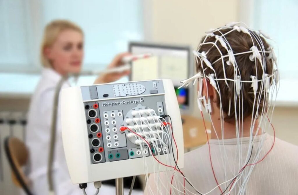
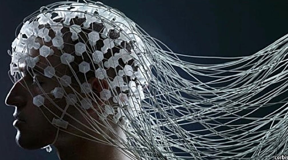
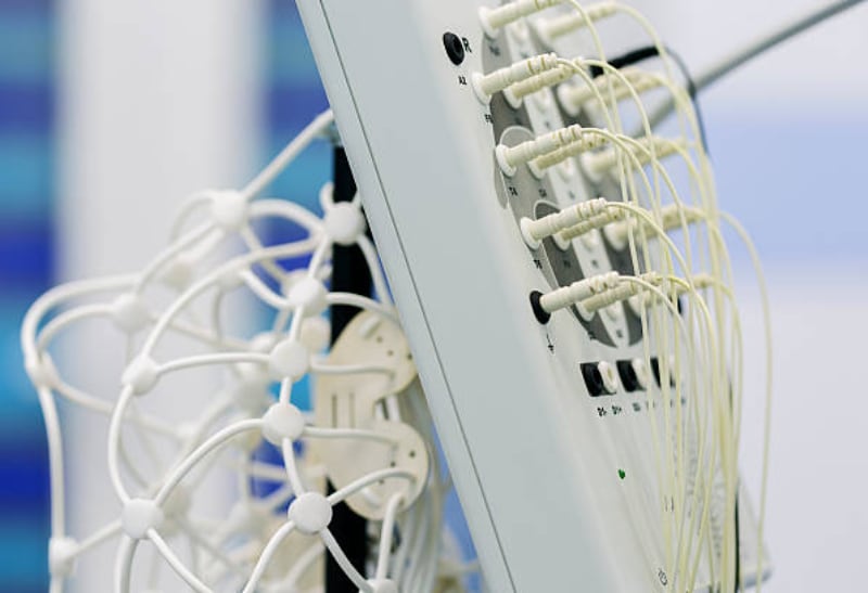
During an EEG, small metal discs called electrodes are attached to the scalp with a special adhesive. These electrodes detect the electrical activity of the brain and transmit the signals to a computer, where they are recorded and analysed. The patient may be asked to perform certain tasks or activities during the test to help the doctor evaluate brain function.

Why is an EEG Performed?

EEG testing is often used to diagnose and monitor conditions such as epilepsy, seizures, sleep disorders, and brain injuries. It can also be used to evaluate brain function during surgery or in intensive care settings. EEG testing is safe, non-invasive, and painless.

Type of EEG

Diagnostic EEG and Long-term monitoring for EEG.

Preparing for an EEG

Before an EEG, it is important to follow any instructions provided by your doctor or technician. This may include avoiding certain medications or caffeine, washing your hair the night before the test, and getting a good night’s sleep. You may also be asked to refrain from eating or drinking for a certain period of time before the test.

During the EEG, the patient will lie on a comfortable bed or chair while the technician attaches the electrodes to the scalp. The patient may be asked to close their eyes or perform certain tasks or activities to stimulate brain activity. The test usually lasts between 30 minutes to an hour.

After the EEG, the electrodes will be removed, and the patient can resume their normal activities. The results of the test will be analysed by a neurologist or other trained healthcare provider, who will provide a diagnosis and treatment plan if necessary.

How to Prepare for EEG

  1. Follow your doctor’s instructions: Your doctor or technician will provide you with instructions on how to prepare for the EEG. It’s essential to follow these instructions carefully to ensure that the test results are accurate. You may be asked to avoid certain medications or caffeine, wash your hair the night before the test, or get a good night’s sleep. Be sure to ask any questions if you’re unsure about anything.
  2. Avoid hair products: Avoid using any hair products such as gel, mousse, hairspray, or conditioner on the day of the test. These products can interfere with the electrodes’ ability to stick to your scalp.
  3. Dress comfortably: Wear comfortable clothing that’s easy to remove and won’t interfere with the electrodes. It’s best to wear a shirt with buttons or a zipper, so you can easily remove it.
  4. Eat and drink as normal: You can eat and drink as normal before the test. However, you may be asked to avoid caffeine or certain foods that can affect brain activity.
  5. Arrive on time: Be sure to arrive on time for your appointment. If you’re late, it may delay the test or cause the appointment to be rescheduled.
  6. Bring a list of medications: Bring a list of all the medications you’re currently taking, including over-the-counter medications and supplements. Your doctor needs to know this information to ensure that the test results are accurate.

How does EEG help?

Here are some ways that EEG can help in the diagnosis of neurological conditions.

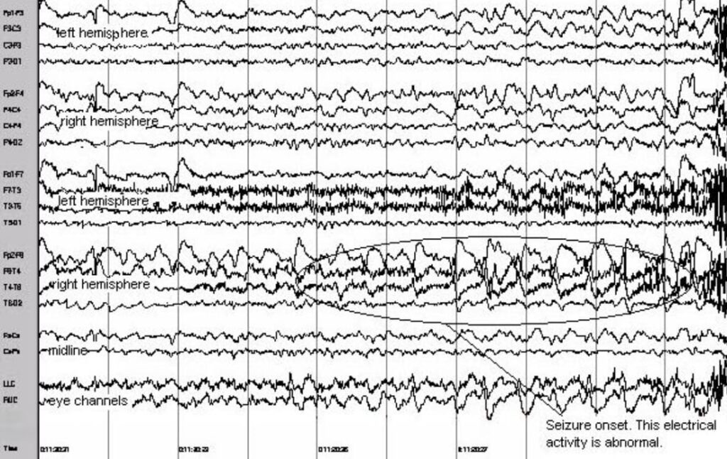
Epilepsy diagnosis: EEG is often used to diagnose epilepsy and seizure disorders. Abnormal electrical activity in the brain can be detected by EEG during a seizure, which can help confirm a diagnosis of epilepsy.

Sleep disorder diagnosis: EEG can also be used to diagnose sleep disorders such as sleep apnea and insomnia. During an EEG sleep study, brain activity is monitored while the patient is sleeping to detect any abnormalities.

Brain injury diagnosis: EEG can be used to diagnose and monitor brain injuries such as concussions and traumatic brain injuries. EEG can detect abnormal electrical activity in the brain that may be indicative of an injury.

Migraine diagnosis: EEG can help diagnose and monitor migraines. During an EEG, changes in brain activity can be detected that may be associated with migraine attacks.

Neurological disorder diagnosis: EEG can help diagnose a range of neurological disorders such as Alzheimer’s disease, Parkinson’s disease, and multiple sclerosis. EEG can detect abnormal electrical activity in the brain that may be indicative of these conditions.

Cost

Fees for EEG at Gippsland Specialist Clinic is $ 250, Medicare rebate is available with minimum of $ 110.00.

How is the Monitoring EEG performed?

Preparation: Before the EEG monitoring begins, the patient’s hair is typically washed to remove any oil or dirt that may interfere with the electrode placement. Any hair products, such as gels or sprays, should also be avoided as they may interfere with the EEG signals.
Electrode placement: EEG electrodes are then placed on the scalp using a special adhesive. The electrodes are typically placed in a specific pattern or montage depending on the type of EEG study being performed. The number and placement of the electrodes can vary depending on the specific needs of the patient.
Recording: Once the electrodes are in place, the EEG is recorded using a computer or other recording device. During the recording, the patient may be asked to perform specific tasks or activities, such as deep breathing or flashing lights, to stimulate brain activity.
Interpretation: The EEG recording is then interpreted by a healthcare provider, such as a neurologist or neurophysiologist, who will analyze the EEG signals and look for any abnormalities or patterns that may indicate a neurological condition.
Follow-up: After the EEG monitoring is complete, the healthcare provider will discuss the results with the patient and develop a treatment plan if necessary. Additional follow-up testing may also be recommended, depending on the specific needs of the patient.

How is the sleep deprived EEG performed?

Sleep deprivation: The patient is asked to stay awake for a specific period before the EEG test. The exact duration of sleep deprivation may vary, but it is typically between 24-48 hours. The patient is advised to avoid caffeine, napping, and other activities that could interfere with sleep.
Electrode placement: EEG electrodes are placed on the scalp using a special adhesive. The electrodes are typically placed in a specific pattern or montage depending on the type of EEG study being performed.

Baseline EEG: Before inducing sleep, a baseline EEG is recorded to establish the patient’s normal brain activity. The patient is asked to relax and remain still during the recording.

Sleep induction: The patient is then asked to relax and attempt to fall asleep. This is typically done in a quiet and darkened room to minimize distractions. The EEG recording is continued throughout the sleep induction process to monitor changes in brain activity.
Stimulation: If necessary, the healthcare provider may use stimulation techniques, such as flashing lights or hyperventilation, to further enhance brain activity during the EEG recording.
Interpretation: After the sleep-deprived EEG is complete, the EEG recording is analysed by a healthcare provider, such as a neurologist or neurophysiologist. The provider will look for any abnormalities or patterns in the EEG signals that may indicate a neurological condition.